


Shoulder Instability Treatment in Nebraska
At Nebraska Hand & Shoulder, we specialize in treating shoulder instability with tailored approaches for every patient. Our expert orthopaedic surgeon offers both surgical and nonsurgical treatments to address your unstable shoulder, aiming to alleviate pain and swelling associated with shoulder joint issues. Trust us to provide the right care for your needs. Schedule an appointment today and discover the Nebraska Hand & Shoulder Institute advantage in Nebraska.
Understanding Shoulder Instability
Diagnosing shoulder instability involves taking a detailed history, using necessary diagnostics like x-rays, and conducting a thorough physical exam. Typically, instability is linked to trauma, with a clear history of a physical incident where the shoulder is painfully dislocated, needing strong sedation in the emergency room or an operating room to reposition it.
Most people who think they've dislocated their shoulder have just hurt the small joint at the top of the shoulder. When we talk about shoulder instability, we are referring to the main glenohumeral joint. There is really no ball-and-socket. The ball is stabilized by ligaments and the overlying muscles.
A strong rotator cuff stabilizes the shoulder, while larger muscles like the deltoid and latissimus dorsi create powerful movements. For smooth shoulder motion, the rotator cuff must work with these bigger muscles to avoid pain through the full range of motion.
Shoulder instability symptoms occur with activity of the shoulder, unlike symptoms from advanced arthritis or rotator cuff tendinitis or impingement, which may be painful even at rest.
X-ray Findings
X-rays at the time of presenting to the emergency room will show the main two bones of the shoulder not aligned properly. Sometimes, a strong shoulder dislocation at the front can occur. After several dislocations, a person might develop a defect in the back of the shoulder ball, called a Hill-Sach's lesion, which is a dent or a chipped piece of bone.
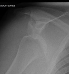
Dislocated Shoulder
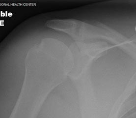
After Reduction
The Hill-Sach’s lesion makes the shoulder more unstable and prone to dislocation, causing apprehension when the shoulder is abducted and externally rotated. This aligns with the apprehension test, which involves moving the arm into the throwing position where dislocation is most likely.
Treatment for Shoulder Instability
At Nebraska Hand & Shoulder, we offer specialized treatment options for those dealing with shoulder instability. Our approach varies based on whether it's a first-time dislocation or a case of chronic shoulder instability.
For first-time dislocations, especially in individuals over 25, we often recommend a conservative approach. This might include using a sling and providing pain relief, along with consultations with a physical therapist to initiate strengthening exercises. After a few weeks, patients can resume activities, avoiding positions that might risk another dislocation.
If shoulder instability persists, we consider surgical treatment options. Our team evaluates the patient’s medical history and decides whether to proceed with a minimally invasive arthroscopic procedure or an open surgery for shoulder dislocation to reconstruct the soft tissue in the glenoid labrum. These ligaments are crucial for preventing future dislocations. The recovery time after surgery can take three to six months, during which physical therapy plays a vital role in ensuring complete healing and restoring shoulder function.
At Nebraska Hand & Shoulder, we strive to provide effective and personalized treatment plans for each patient, focusing on surgical treatment coupled with physical therapy to optimize recovery outcomes.
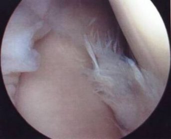
Labral Tear Diagnosis
There is significant debate surrounding the diagnosis of a torn glenoid labrum, which is a rare injury. In older individuals, degenerative tears may be asymptomatic but still show up on an MRI scan.
However, many MRI interpretations suggesting a torn labrum are incorrect. In fact, during surgery, surgeons often find the labrum to be intact, and the tear may not be related to the patient's symptoms. Labral tears are typically categorized as degenerative or traumatic, repairable or non-repairable.
Individuals over 40 are generally not candidates for repair, and instead, the torn portion is trimmed if it causes symptoms. Reattachment is necessary for labral tears torn off the glenoid rim to prevent instability and pain, although this type of tear is infrequent.
Contact Nebraska Hand & Shoulder for Expert Care
If you have chronic shoulder instability, contact Nebraska Hand & Shoulder for specialized treatment. Our team uses advanced diagnostics, like imaging tests, to accurately assess your shoulder.
Whether you need nonsurgical treatments or surgery, our experts are committed to restoring your shoulder's function and improving your quality of life. Schedule your consultation today and take the first step towards effective shoulder instability management.