Tarsal Tunnel Syndrome

Introduction 

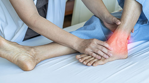
Tarsal tunnel syndrome (TTS) was first reported in 1962 by Drs. Keck and Lam. It relates to symptoms developing because of nerve entrapment at the ankle. There is an anterior (top of foot) and posterior (bottom of the foot) tarsal tunnel. Different nerves provide sensibility (carry sensation) from each. Symptoms include tingling and numbness of the feet involving some or all of the toes or limited to the heel. Pain about the foot and/or ankle and a feeling of tightness about the ankle are also typical symptoms.

Pain in diabetics is usually described as burning and may involve the whole foot and calf i.e:'stocking'' due to associated peroneal nerve entrapment at the knee.

Symptoms may be worse at night when one is trying to lie down and may feel better getting up and walking on the feet though prolonged walking is often not tolerated and, in fact, walking duration may be limited. Because of the discomfort, a person may be taking anti­ inflammatory medication in the futile hope that this will treat the "arthritis", which is the wrong diagnosis. Tarsal tunnel syndrome occurs at increased frequency in diabetics causing decreased sensibility. This is associated with a high rate of skin ulceration and ultimately toe or foot amputation when the ulcers become infected and fail to heal.

Aggressive treatment of the tarsal tunnel syndrome may eliminate the abnormal sensation in the feet and prevent skin breakdown!

Download our Tarsal Tunnel Patient Brochure

tarsal tunnel syndrome

Who Gets Tarsal Tunnel Syndrome?

This is a problem restricted to adults usually beyond the age of 30. No absolute cause is usually identifiable though obesity and diabetes are frequently associated. In diabetics, increased fluid is accumulated in nerves because of the accumulation of non-digestible sugar sorbitol. There may also be thickening of the nerve from amyloidosis. Tarsal tunnel syndrome may develop even with well­ controlled blood sugar. The symptoms are frequently misconstrued as "diabetic neuropathy" which is an untreatable disease-specific entity but tarsal tunnel syndrome is, in fact, correctible. As in carpal tunnel syndrome (CTS), tarsal tunnel syndrome (TIS) occurs much more commonly in women.

Painful heel syndrome usually occurs in markedly overweight people and is sometimes due to nerve entrapment at the tarsal tunnel rather than "plantar fasciitis", another common problem.

What Causes TTS?

The culprit for a posterior tarsal tunnel is pressure upon the tibial nerve going from the calf around the inner anklebone and then through the fibrous abductor hallucis muscle origin (muscle to the great toe). Two branches-the calcaneal and the plantar-have been associated with ITS. Age, physical condition, and sometimes even a prior injury such as a broken ankle may be related. In anterior tarsal tunnel the deep peroneal nerve may be compressed beneath the inferior extensor retinaculum on the front side of the ankle. This causes pain over the anterior ankle and decreased sensibility or burning in the space between the big toe and the second toe. A feeling of tightness about the ankle may exist. In diabetics common peroneal nerve entrapment just below the knee often occurs at the same time. Symptoms of common persistent entrapment may overlap or be masked by entrapment of the tibial nerve at the tarsal tunnel. Preoperative nerve testing with the Pressure Specified Sensory DeviceTM helps to identify single or multiple nerve involvement and tends to be useful for monitoring nerve recovery. In diabetics developing TTS is probably related to subtle nerve enlargement in confined spaces as well as other physiologic changes.

Diabetics and Peripheral Neuropathy 

Peripheral neuropathy means malfunctioning of the nerves outside of the brain and spinal cord that serve to activate your muscles and provide sensibility to the feet and hands. The term includes entrapment neuropathy such as carpal tunnel syndrome (CTS), cubital tunnel syndrome (CTS), and tarsal tunnel syndrome (TTS).

Peripheral neuropathy also includes generalized nerve decay such as diabetic neuropathy, chemical neuropathy, alcoholic neuropathy, etc. The distinction is important because there is very little treatment available for non-entrapment neuropathy, diabetic neuropathy, and alcoholic neuropathy, while entrapment neuropathies such as CTS and TTS if accurately diagnosed and treated before irreversible damage has occurred generally result in very good symptom relief.

Non-surgical treatment is centered around hiding symptoms with medications that interrupt or reduce nerve activity or reduce the perception of nerve activity such as Lyrica (pregabalin), amitriptyline and its relatives, and Tramadol. Only entrapment neuropathy generally has a good prognosis with predictable outcomes. Of particular importance in this context is the misunderstanding with regard to diabetic peripheral neuropathy.

A high percentage of diabetics develop a nerve disorder. That makes the nerves more sensitive to external pressure. Therefore, it is very common for a diabetic to also develop entrapment neuropathy. Many of the diabetic symptoms in the hands and feet are related to superimposed nerve entrapment and thus a treatable nerve entrapment such as carpal, cubital tunnel syndrome, and tarsal tunnel syndrome needs to be excluded before condemning the diabetic person to a life of pain, numbness and decreased ability to use the hand or walk from an untreatable nerve process. Decompressing diabetic peripheral nerves has been shown to have almost equal results to the nondiabetic if patients are carefully scrutinized. An unexpected benefit of decompression of the tibial and fibular nerves affecting the feet in diabetics is that of prevention of ulceration and healing of ulcers already present in selected patients with diabetes.

To make this possible we have the first and only Pressure Specified Sensory Device in Nebraska invented by Dr. A Lee Dellon.

Dr. Ichtertz visited with Dr. Dellon, Professor of Plastic Surgery and Neurosurgery at John Hopkins University Medical Center and author of over 350 scientific articles. The goal was to gain a full understanding of the use of the Pressure-Specified Sensory Device (PSSD). Dr. Ichtertz has utilized this technology to further enhance the diagnosis and improve the treatment of ulnar nerve entrapment, both nonoperative and operative in diabetes and diabetics alike.

Many people diagnosed with “plantar fasciitis" actually have entrapment of a branch of the tibial nerve as it enters the heel hence the treatment of plantar facial release which actually decompresses the nerve branch. Usually, before offering surgical release for a painful heel thought to be plantar fasciitis, stretching, shoot inserts to take direct pressure off of the heel and a cortisone injection are tried first.

How is the diagnosis confirmed?

Symptoms about the foot and ankle require careful tests for a malfunctioning nerve. For example, tapping upon the tibial nerve at the ankle may result in local pain or tingling (Tinel's sign) or that is referred to the toes in an "electric shock" fashion. Though it's more difficult to identify, there may be weakness of great toe movement away from the foot (abductor hallucis muscle weakness).

Absolute confirmation of nerve dysfunction is obtained with a thorough nerve conduction study or the newly introduced quantitative sensory test (Pressure­ Specific Sensory Device™).

This is a reliable, non­ invasive (painless), computer­ based technique of identifying and tracking subtle changes in sensory function. A conventional nerve conduction study (NCS) is advisable to localize nerve entrapment. EMO (poking the muscles with needles to evaluate electrical activity) is still sometimes necessary. Peroneal nerve entrapment at the fibular neck Gust below the knee) may result in local discomfort just below the outer knee. Though it usually results in numbness or tightness about the ankle and the top of the foot. If it goes untreated foot drop, and inability to lift one's foot, may develop.

IBM used for testing tarsal tunnel syndrome

How is TTS treated?

In people who are excessively overweight (medically known as morbid obesity) drastic weight loss may be of value both in eliminating the problem and in preparing for surgery. Optimizing serum glucose levels in a diabetic or use of an arch support in a person with arch-related discomfort may benefit others. Ultimately, most people with TTS will probably require a minor outpatient operation.

This is performed with spinal or general anesthesia though it's also possible under heavy intravenous sedation plus local anesthesia.

Walking upon the foot is not absolutely contraindicated as long as it's kept covered enough for the wound to seal itself. Avoiding excessive walking is also advisable. A cane held in the opposite hand or crutches may be used for several weeks It does take about 2 to 3 weeks before sutures are ready for removal and ultimately, I expect someone to be completely healed within about 6-8 months or less.

Marked relief of pain is often possible shortly after the surgery. People typically realize benefit within 24 hours of the operation though it may take longer depending upon the magnitude and character of one's symptoms.

Early treatment before irreversible nerve damage yields the best results.

Diabetics with TIS have an enormous potential gain: improved comfort, healing of open ulcers, and prevention of sores/ulcers has been demonstrated for years by A. Lee Dellon, M.D., Professor of Plastic Surgery and Professor of Neurosurgery at Johns Hopkins University Medical Center. Drs. Wieman and Patel at the Department of Surgery in Louisville, Kentucky, and Dr. Caffee at the University of Florida, Department of Surgery and others also report good results.

Who should be tested?

The American Diabetes Association recommends quantitative sensory (PSSD) testing of the feet of diabetics once a year. The person suited for intervention is beginning to experience loss of sensibility, but their feet are not yet "numb". The greater the sensory loss, the greater the nerve cell (axon) loss. PSSD enables the doctor to monitor gradual changes and discern which nerves are involved early enough to correct the problem and prevent skin breakdown.

Results/Benefits

For diabetics who meet criteria for surgery, 60% to 80% or 6 to 8 out of 10 people, get a good result according to Dr. Dellon. Drs.

Wieman and Patel reported success in 92% (24 of 26) if the patients had electrical sensation upon tapping the involved nerve pre op (Tinel's sign). Dr. Caffee reported 86% good results. Variation in outcomes reflect patient differences and variation in delay to surgery. The majority of patients note almost immediate elimination of burning pain after nerve decompression. Most notable has been the healing of long-term ulcers upon improvement in nerve function.

Outcome/benefit from surgery?

Recovery rate varies. For the first couple of weeks, the patient is advised to elevate the foot on a pillow above heart level to reduce or prevent throbbing related to swelling. If anti-inflammatory medication is not contraindicated, its use will markedly improve one's comfort level. Within 2-5 days most people are back to sedentary jobs.

References

Borges, LF, Hallette, M, Selkoe, DJ, Welch, K, "The anterior tarsal tunnel syndrome." J Neurosurg, 1981; 54: 89-92

Dellon, AL, "Computer-assisted sensibility evaluation and surgical treatment of the tarsal tunnel syndrome." Advances in Podiatric Medicine and Surgery, 1996; 2: 17.

Gessini, L, Jandolo, B, Pietrangeli, A, "The anterior tarsal syndrome." JBJS, 1984; 66A: 786-787.

Kopell, HP, Thompson, WAL, "Peripheral entrapment neuropathies of the lower extremity." N Engl J Med, 1960; 262: 56-60.

Marinacci, AA, "Neurological syndromes of the tarsal tunnels." Bull LA Neurol Soc, 1968; 33: 90-100.

Bailie, OS, Kelikian, AS, "Tarsal tunnel syndrome: Diagnosis, surgical

technique, and functional outcome." Foot & Ankle International, 1998; 19: 65-72.

Caffee, HH, "Treatment for diabetic neuropathy by decompression of the posterior tibial nerve." Plast & Reconst Surg, 2000; I06: 8 I 3.

Dellon, AL, "Computer-assisted sensibility evaluation and surgical treatment of the tarsal tunnel syndrome." Advances in Podiatric Medicine and Surgery, 1996; 2: 17.

Keck, C, "The tarsal tunnel syndrome." JBJS. 1962; 44: I 80-182. 

Lam, SJS, "A tarsal tunnel syndrome." Lancet, 1962; 2: 1354-1355.

Wieman, TJ, Patel, VG, "Treatment ofhyperesthetic neuropathic pain in

diabetics." Annals of Surg, 1998; 221: 660-665.

Baxter, DE, Pfeffer, GB, Thigpen, M, "Chronic heel pain: Treatment rationale." Orthop Clin North Am, 1989; 20: 563-569.

Baxter, DE, Thigpen, CM, "Heel pain-operative results." Foot and Ankle, I 984; 5: I: 16.

Beskin, JL, "Nerve entrapment syndromes of the foot and ankle." J Am Acad Orthop Surg; 1997: 5: 261-269.

Przylucki, H, Jones, CL, "Entrapment neuropathy of muscle branch lateral plantar nerve." J Am. Podiatrc Assoc, 1981; 71: 3: I 19.

Dellon, AL, "Deciding when heel pain is of neural origin." Journal Foot & Ankle Surgery, 200 I ;40: 5: 341-345.

Galardi, G, Amadio, S, Maderna, L, Meraviglia, MV, Bruanti. L, Conte, GD,

Comi, G, "Electrophysiologic studies in tarsal tunnel syndrome." Am J Phys Med RehabiI, 1994, 76: 193-198.

Saeed, MA, Gatens, PF, "Compound nerve action potentials of the medial and lateral plantar nerves through the tarsal tunnel." Arch Phys Med Rehabil, 1982; 63: 304.

Tassler, PL, Dellon, AL, "Correlation of measurements of pressure perception using the Pressure-Specified Device with electrodiagnostic testing." JOEM, 1995; 37: 862-866.

Tassler, PL, Dellon, AL, "Pressure perception in the normal lower extremity and in tarsal tunnel syndrome." Muscle & Nerve, 1996; 19: 285-289.

Tassler, PL, Dellon, AL, Scheffler, NM, "Computer-assisted measurement in diabetic patients with and without foot ulceration." J Am Podiatric Med Assoc, 1995;85: II: 679-684.

References

Anderson, Bruce Carl, et al, “Treatment of de Quervain’s tenosynovitis with corticosteroids.” Arthritis and Rheumatism, Vol. 34, No. 7, July 1991, pp. 793-798. Badalamente, M.A., “Pathobiology of the Human A1 Pulley in Trigger Finger.” The Journal of Hand Surery, Vol. 16A, 1991, pp. 714-721. Bishop, A.T., “Extensor Triggering in de Quervain’s Stenosing Tenosynovitis.” The Journal of Hand Surgery. Vol. 24A, 1999, pp. 1311-1314. Dinham, J.M., Meggitt, B.F., “Trigger thumbs in children: a review of the natural history and indications for treatment in 105 patients.” Journal of Bone and Joint Surgery, Vol. 56B, No 1, 1974, pp. 153-155. Doyle, J.R., Blythe, W.F., “The finger flexor tendon sheath and pulley: anatomy and reconstruction.” AAOS Symposium on Tendon Surgery in the Hand, St. Louis, 1975. The C.B. Mosby Company, pp. 81-87. Doyle, J.R., “Anatomy of the Flexor Tendon Sheath and Pulleys of the Thumb.”, The Journal of Hand Surgery, Vol. 2, 1977, pp. 149-151. Eastwood, D.M., Gupta, K.J., Johnson, D.P., “Percutaneous Release of Trigger Finger: An Office Prodedure.” Journal of Hand Surgery, Vol. 17A, 1992, pp. 114-117. Ezaki, M., “Trigger Finger in Children.”, The Journal of Hand Surgery, Vol. 24A, 1999, pp. 1156-1161. Fulcher SM, Hill, J.J.; “An Analysis of Patients with Multiple Trigger Digits.” [Poster] 56th Annual Meeting of the American Society for Surgery of the Hand Oct. 4, 2001, Seattle, WA. Gray, R.G. and Gottlieb, N.L., “Hand flexor tenosynovitis in rheumatoid arthritis: prevalence, distribution, and associated rheumatic features.” Arthritis and Rheumatism, Vol. 20, No. 4, 1970, pp. 103-108. Griggs, S.M., “Treatment of Trigger Finger in Patients with Diabetes Mellitus.” The Journal of Hand Surgery, Vol. 20, 1995, pp. 787-789. Harvey, Francis J., et al, “deQuervain’s disease: surgical and non-surgical treatment.” Journal of Hand Surgery, Vol. 15A, No. 1, January 1990, pp. 83-87. (Diagram 4). Hoffmann, R., “Open Versus Percutaneous Release of the A1-Pulley for Stenosing Tendovaginitis: A Prospective Randomized Trial.”, Techniques in Hand & Upper Extremity Surgery, Vol. 12, 2008, pp. 183-187. Hollander, J.L., et al, “Hydrocortisone and cortisone injected into arthritic joints, comparative effects of and use of the hydrocortisone as a local anti-arthritic.” JAMA, Vol. 147, 1951, pp. 1629-1635. Jackson, W.T., et al, “Anatomical variations of the first extensor compartment of the wrist: a clinical and anatomical study.” Journal of Bone and Joint Surgery, Vol. 68A, No. 6, July 1986, pp. 923-926. Lane, L.B., “Commentary: Percutaneous Release of the First Annular Pulley.” The Journal of Hand Surgery, Vol. 20, 1995, pp. 785-786. Lapidus, P.W., “Stenosing tenovaginitis.” Surgical Clinics of North America, Vol. 33, 1953, pp. 1317*1347. Lubahn, J., “Complications of Open Trigger Finger Release.”, The Journal of Hand Surgery, Vol. 35A, 2010, pp. 594-596. Medl, W.T., “Tendonitis, tenosynovitis, trigger fingers, and deQuervain’s disease.” Orthopaedic Clinic of North America, Vol. 1, No. 2, 1970, pp. 375-382. Otto, N., Wehby, M., et al, “Steroid injections for tenosynovitis in the hand.” Orthopaedic Review, Vol. 15, No. 5, May 1986. Patel, M.R., Bassini, L., “Trigger fingers and thumb: when to splint, inject or operate” Journal of Hand Surgery Vol. 17A, No. 1, Jan. 1992, pp. 110-113. Patel, M.R., “Percutaneous Release of Trigger Digit With and Without Cortisone Injection.” The Journal of Hand Surgery, Vol. 22A, 1997, 150-155. Peimer, C.A., “Release of the Sixth Dorsal Compartment.” The Journal of Hand Surgery, Vol. 19A, 1994, pp. 599-601. Pratt, H.L., Bunnell, S., “Use of compound F (hydrocortisone in op) in operative and non-operative conditions of the hand.” Journal of Bone and Joint Surgery, Vol. 35, 1953, pp. 94-102. Roth, J.H., “Percutaneous A1 Pulley Release: A Cadaveric Study.” The Journal of Hand Surgery, Vol. 20A, 1995, 781-784. Sampson, S.P., et al, “Pathobiology of human A-1 pulley in trigger finger.” Journal of Hand Surgery, Vol. 16A, 1991, pp. 714-721. Tanaka, J., “Percutaneous Trigger Finger Release.”, Techniques in Hand and Upper Extremity Surgery, Vol. 3, 1999, pp. 52-57. Weilby, A., “Trigger finger: incidence in children and adults and the possibility of a predisposition in certain age groups.” Acta Orthopaedics Scandinavia, Vol. 41, 1970, pp. 419-427. Weiss, A.C., “Treatment of De Quervain’s Disease.” The Journal of Hand Surgery, Vol. 19A, 1994, pp. 595-598. Witczak, J.W., “Triggering of the Thumb with de Quervain’s Stenosing Tendovaginitis.” The Journal of Hand Surgery, Vol. 15, 1990, pp. 265-268. Witt,J. “Treatment of de Quervain Tenosynovitis: A Prospective Study of the Results of Injection of Steroids and Immobilization in a Splint.” Journal of Bone & Joint Surgery, Vol. 73, 1991, pp. 219-222 Woods, T.H.E., “deQuervain’s disease: a plea for early operation: a report on 40 cases.” British Journal of Surgery, Vol. 51, No. 5, May 1964, pp. 358-359.